ventricle
US /ˈvɛntrɪkəl/
・UK /ˈventrɪkl/
C2
n.NounChamber of the heart that pumps blood to arteries
Fatty diets can damage one's ventricle and cause heart complications
Video subtitles
Watch a Transcatheter Aortic Valve Replacement (TAVR) Procedure at St. Luke's in Cedar Rapids, Iowa

- The aortic valve is the valve that sits between the left bottom chamber of the heart, we call it left ventricle, which is the main chamber of the heart that pumps the blood to the whole body, and between the aorta, which is the blood vessel, the largest blood vessel that we have, that comes out of this left ventricle and distributes the blood to the whole body.
The aortic valve is the valve that sits between the left bottom chamber of the heart, we call it left ventricle, which is the main chamber of the heart that pumps the blood to the whole body, and between the aorta, which is the blood vessel, the largest blood vessel that we have, that comes out of this left ventricle and distributes the blood to the whole body.
- The aortic valve is the valve that sits between the left bottom chamber of the heart—we call it left ventricle—which is the main chamber of the heart that pumps the blood to the whole body, and
The aortic valve is the valve that sits between the left bottom chamber of the heart—we call it left ventricle—which is the main chamber of the heart that pumps the blood to the whole body, and
Exploring the Heart - The Circulatory System!

- through the second room called the right ventricle, and through the second door called the pulmonary valve.
through the second room called the right ventricle, and through the second door called the pulmonary valve.
- and through the second room called the "right ventricle",
and through the second room called the "right ventricle",
Anatomy and Physiology of the Cardiovascular System: Heart Anatomy
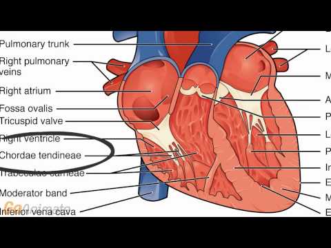
- chamber called the right ventricle. The blood flows past a valve called the tricuspid
chamber called the right ventricle. The blood flows past a valve called the tricuspid
- Blood flows from the right atrium to a larger chamber called the right ventricle.
Blood flows from the right atrium to a larger chamber called the right ventricle.
Shashank Sinha, M.D. | Evaluation and Management of Cardiogenic Shock in 2025

- She's noted to have minimal pulsatility of her LV, a severely dilated left ventricle of 9 centimeters, and a dilated and moderately dysfunctional right ventricle.
She's noted to have minimal pulsatility of her LV, a severely dilated left ventricle of 9 centimeters, and a dilated and moderately dysfunctional right ventricle.
- She's noted to have minimal pulsatility of her LV a severely dilated left ventricle of 9 cm and a dilated and moderately dysfunctional right ventricle and an impella.
She's noted to have minimal pulsatility of her LV a severely dilated left ventricle of 9 cm and a dilated and moderately dysfunctional right ventricle and an impella.
Cardiovascular | Anatomy of the Heart | Heart Model
- It's what takes deoxygenated blood from the right ventricle to the lungs to get oxygenated.
It's what takes deoxygenated blood from the right ventricle to the lungs to get oxygenated.
- It's what takes deoxygenated blood from the right ventricle to the lungs to get oxygenated.
It's what takes deoxygenated blood from the right ventricle to the lungs to get oxygenated.
Apr 03 2026 This Week in Cardiology

- This is a device placed in the left ventricle, and it provides liters of cardiac output.
This is a device placed in the left ventricle, and it provides liters of cardiac output.
- This is a device placed in the left ventricle, and it provides liters of cardiac output.
This is a device placed in the left ventricle, and it provides liters of cardiac output.
16) Pharmacology of vasoactive therapies used in low-output heart failure and cardiogenic shock.
- it's, it's really the left ventricular outflow tract obstruction or, or even the severe aortic stenosis patients pre-TAVR, um, where essentially the fixed cardiac output pushing O2-rich blood through a pinhole, um, you need systemic vasoconstriction in order to maintain a blood pressure in those patients because increasing inotropy is not gonna make more blood be ejected from the left ventricle, pushing it through a pinhole.
it's, it's really the left ventricular outflow tract obstruction or, or even the severe aortic stenosis patients pre-TAVR, um, where essentially the fixed cardiac output pushing O2-rich blood through a pinhole, um, you need systemic vasoconstriction in order to maintain a blood pressure in those patients because increasing inotropy is not gonna make more blood be ejected from the left ventricle, pushing it through a pinhole.
- it's, it's really the left ventricular outflow tract obstruction or even the severe aortic stenosis patients pre-TAVR, um, where essentially the fixed cardiac output pushing O2-rich blood through a pinhole, um, you need systemic vasoconstriction in order to maintain a blood pressure in those patients because increasing inotropy is not gonna make more blood be ejected from the left ventricle, pushing it through a pinhole.
it's, it's really the left ventricular outflow tract obstruction or even the severe aortic stenosis patients pre-TAVR, um, where essentially the fixed cardiac output pushing O2-rich blood through a pinhole, um, you need systemic vasoconstriction in order to maintain a blood pressure in those patients because increasing inotropy is not gonna make more blood be ejected from the left ventricle, pushing it through a pinhole.
Chapter 6 - The Cardiorespiratory, Endocrine, and Digestive Systems
- And if we go back, well the same, it's the same picture, but you know, right atrium, right ventricle, left atrium, left ventricle, and they are going to be working independently right and left sides.
And if we go back, well the same, it's the same picture, but you know, right atrium, right ventricle, left atrium, left ventricle, and they are going to be working independently right and left sides.
- if we go back, well, the same it's the same picture, but, you know, right atrium, right ventricle, left atrium, left ventricle,
if we go back, well, the same it's the same picture, but, you know, right atrium, right ventricle, left atrium, left ventricle,
How I Take Notes with My iPad Pro in Lectures (Notability & GoodNotes) + Free Template

- Why are the walls of the left ventricle much thicker than the walls of the right ventricle?
Why are the walls of the left ventricle much thicker than the walls of the right ventricle?
- What on earth is a ventricle?
What on earth is a ventricle?
ACC 26: The STEMI-DTU Trial – Primary Unloading and Delayed Reperfusion in STEMI

- Another important aspect for clinicians to understand from this trial, in contemporary clinical practice with a door-to-balloon time of 54 minutes, excellent execution of anterior STEMI management, over 30% of the left ventricle was left damaged by a heart attack.
Another important aspect for clinicians to understand from this trial, in contemporary clinical practice with a door-to-balloon time of 54 minutes, excellent execution of anterior STEMI management, over 30% of the left ventricle was left damaged by a heart attack.