Subtitles & vocabulary
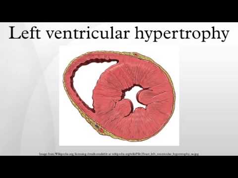
Left ventricular hypertrophy
00
Ting Huang posted on 2016/05/17Save
Video vocabulary

lead
US /lid/
・
UK /li:d/
- Noun (Countable/Uncountable)
- Wire for electricity, computer, etc.; cable
- Information that could help to solve a crime
- Adjective
- Being the main part in movies or plays
A1TOEIC
More pressure
US /ˈprɛʃɚ/
・
UK /'preʃə(r)/
- Noun (Countable/Uncountable)
- Anxiety caused by difficult problems
- Force, weight when pressing against a thing
- Transitive Verb
- To apply force to something
- To persuade or force someone to do something
A2TOEIC
More disease
US /dɪˈziz/
・
UK /dɪˈzi:z/
- Noun (Countable/Uncountable)
- Illness that affects a person, animal, or plant
- A disorder of structure or function in a plant, especially one caused by a pathogen.
- Transitive Verb
- To affect with disease; to corrupt or sicken.
A2TOEIC
More measure
US /ˈmɛʒɚ/
・
UK /ˈmeʒə(r)/
- Noun (Countable/Uncountable)
- Plan to achieve a desired result
- Tool used to calculate the size of something
- Transitive Verb
- To determine the value or importance of something
- To calculate size, weight or temperature of
A1TOEIC
More Use Energy
Unlock Vocabulary
Unlock pronunciation, explanations, and filters

