Subtitles & vocabulary
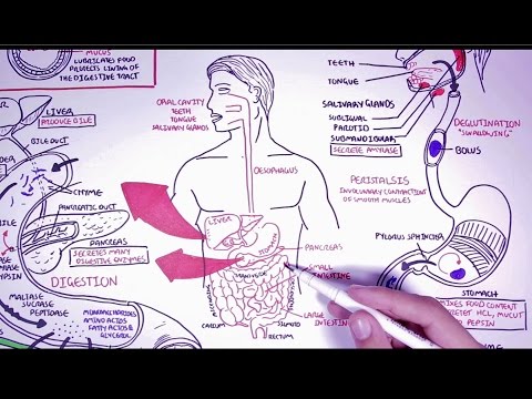
Hungry Microbiome: The Digestive System
00
Lizzy posted on 2015/06/18Save
Video vocabulary

layer
US /ˈleɚ/
・
UK /ˈleiə/
- Noun (Countable/Uncountable)
- One of several sheets of a material or object
- A covering of something spread over a surface.
- Transitive Verb
- To put things one on top of another
B1TOEIC
More system
US /ˈsɪstəm/
・
UK /'sɪstəm/
- Noun (Countable/Uncountable)
- Set of organized, planned ideas that work together
- A set of principles or procedures according to which something is done; an organized scheme or method.
- Adjective
- Working in an organized, logical way
A1TOEIC
More absorb
US /əbˈsɔrb, -ˈzɔrb/
・
UK /əb'sɔ:b/
- Transitive Verb
- To take up all attention / energy of something
- To take in a liquid; soak up
B1TOEIC
More bacteria
US /bækˈtɪriə/
・
UK /bæk'tɪərɪə/
- Noun (plural)
- The very small creatures that can cause disease
B2
More Use Energy
Unlock Vocabulary
Unlock pronunciation, explanations, and filters

